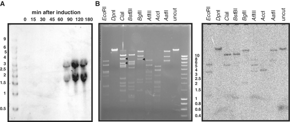
Figure 7.
Detection of transcription start sites in the 80α genome. (A) Northern blot of RNA isolated at the indicated time points after MC induction of an 80α lysogen and probed with a biotinylated terS oligonucleotide. Sizes in kilobase of the markers (Riboladder Long, Bioline) are shown on the left. (B) Agarose gel (left) and Southern blot (right) of 80α virion DNA, digested with the indicated restriction enzymes and probed with total RNA that was isolated from infected cells at various time points, pooled, and 5′-end-labeled at triphosphate termini using vaccinia virus capping enzyme. Sizes in kilobase of the markers (HyperLadder I, Bioline) are indicated to the right of the gel. Black arrowheads on the gel denote examples of unique large fragments from the late region that are not detected in the blot, but to which hybridization would be expected if there were additional transcription start sites downstream of terS.

