Figure 2.
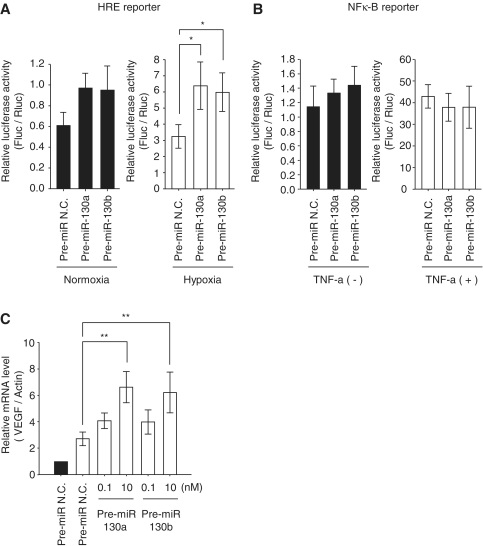
Hypoxia signaling in the miR-130 family. (A and B) Reporter assays with HRE reporter plasmid or NF-κB reporter plasmid are shown. HEK293 cells were transfected with reporter plasmid and pre-miR-130 family members (10 nM). After 48 h transfection Cells were exposed to hypoxia (A, open bars) or 20 ng/ml TNF-α for 12 h (B, open bars). Data are presented as means ± SD (n = 5, *P < 0.01). Solid bars show the luciferase activity without treatment in the pre-miR N.C. and pre-miR-130a family members (solid bars). The P-values in the left of (A) are as follows: Control versus 130a: P = 0.04; control versus 130b: P = 0.02. Firefly luciferase activities were analyzed and corrected for transfection efficiency by the Renilla luciferase activity. (C) In the transient transfection of pre-miRNAs (0.1 and 10 nM), after 48 h cells were exposed to hypoxia, and VEGF mRNA levels were analyzed by qRT–PCR. The mRNA levels were corrected for differences in β-actin mRNA. The values during hypoxia (open bars) are represented as the ratio of control in normoxia (solid bars). Results are presented as means ± SD (n = 6, **P < 0.001). Pre-miR N.C. (10 nM) was used as a control.

