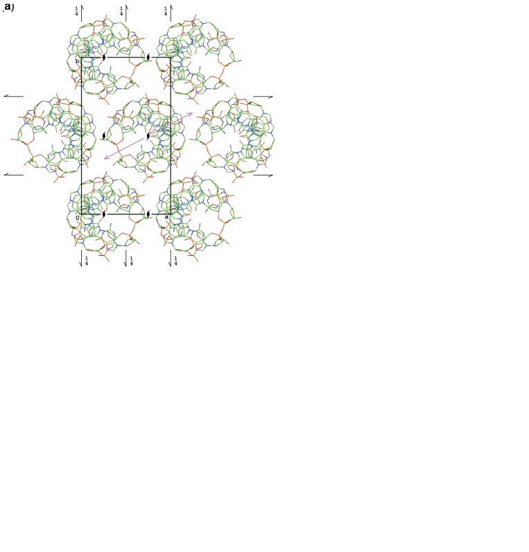
Figure 1.
Packing of the Z-DNA molecules in the current structure and in other structures marked A in Table 1 (a and b), and in the structures marked B (c and d). In (a) and (c) the structures are projected down the crystal a-axis, coincident with the helix axis. In (b) and (d), the structures are projected along the helix 2-fold axes (marked as magenta arrows in a and c), lying in the b and c plane.

