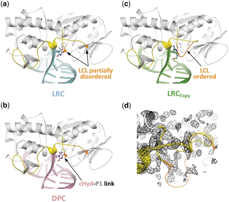
Figure 4.
Overview of the LRC and DPC structures. (a, b and c) Overall structures of Fpg Hyd recognition complex (LRC, blue), Hyd-DNA Fpg covalent complex (DPC, pink) and Fpg FapyG recognition complex (LRCFapy, green, pdbid:1XC8). The ribbon representation of Fpg is in grey and the lesion-capping loop (LCL) is highlighted in yellow. In the structure of LCRFapy, the highly flexible part of LCL is ordered and highlighted in orange. (d) Close-up views of LCL. The partially disordered LCL (thick cylinder) from LRC is superimposed with the full LCL from LRCFapy (thin cylinder).

