Figure 1.
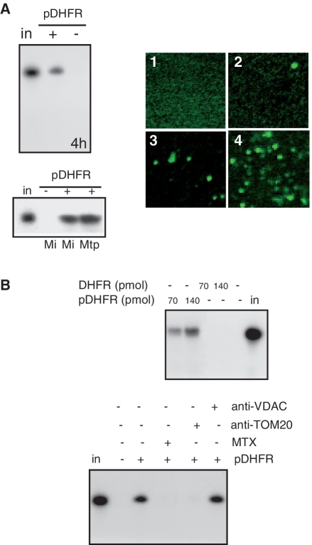
pDHFR increases tRNA import into isolated potato mitochondria. (A) On the left: 32P-labeled in vitro-transcribed tRNAAla was incubated with isolated potato mitochondria (25) in the absence (−) or presence (+) of 35 pmol of pDHFR. Following standard import conditions, RNase treatment was performed either on mitochondria (Mi) or on mitoplasts (Mtp). RNAs were fractionated on a denaturing polyacrylamide gel. Equivalent loading was checked by ethidium bromide staining prior to autoradiography visualization (4 h exposure). On the right: visualization under confocal microscope of Alexa Fluor-labeled in vitro transcribed tRNAAla incubated with isolated potato mitochondria in the absence (2) or presence (3 and 4) of 35 pmol of pDHFR. Visualization was performed after 5 min (3) or 25 min (2 and 4) of incubation. Alexa Fluor-labeled in vitro transcribed tRNAAla in import medium without mitochondria was used as a control (1). (B) 32P-labeled in vitro-transcribed tRNAAla was incubated with isolated potato mitochondria in the absence (−) or presence (70 and 140 pmol) of pDHFR or DHFR. Methotrexate (MTX, 50 nM), or antibodies against VDAC or TOM20 (25) were added to the standard import mixture and incubated for 10 min before adding the labeled tRNAAla. in: 10% of input RNA (2 fmol).

