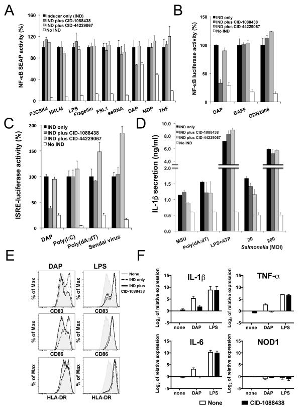
Figure 2. CID-1088438 specifically inhibits NOD1-dependent signaling pathways.
(A) PMA-differentiated THP.1-cells containing NF-κB-driven SEAP (105 cells/well in 96-well plate) were cultured with or without 5 μM CID-1088438 or CID-44229067 and various TLR/NLR inducers: 0.5 μg/ml Pam3CSK4 (TLR1/TLR2), 5×10⁷ cells/ml HKLM (TLR2), 1 μg/ml FSL-1 (TLR6/2), 0.5 μg/ml LPS (TLR4), 0.5 μg/ml Flagellin (TLR5), 1 μg/ml ssRNA40 (TLR8), 5 μg/ml γ-tri-DAP (NOD1), 5 μg/ml MDP (NOD2) or 5 ng/ml TNFα. After 24h incubation, SEAP activity in culture supernatants was measured, expressing data as percentage relative to treatment with inducer only (indicated as 100%; mean ± SEM, n=2). (B) 697 cells stably containing a NF-κB-luciferase reporter gene (105 cells/well in 96-well plate) were cultured with or without 10 μM CID-1088438 or CID-44229067, in combination with 20 μg/ml γ-tri-DAP, 100 ng/ml BAFF or 5 μM ODN2006 (TLR9). Luciferase activity was measured 24h later (mean ± SD; n=3). (C) 293T cells, stably expressing luciferase reporter gene driven by IFN responsive elements (105 cells/well in 96-well plate), were cultured with or without 5 μM CID-1088438 or CID-44229067, in combination with 10 μg/ml γ-tri-DAP (NOD1), 1 μg/ml poly(I:C) with lipid transfection (LyoVec) (RIG-I/MDA-5), 1 μg/ml Poly(dA:dT) (LyoVec) (IRF3) or Sendai Virus (classical IRF3 inducer). Luciferase activity was measured after 24 hrs (mean ± SD; n=4). (D) RAW264.7 cells (5×10⁴ cells/well in 96-well plate) were treated with 5 μM of CID-1088438 or CID-44229067, then stimulated with 100 ng/ml monosodium urate (MSU), 1 μg/ml poly(dA:dT) or 1 μg/ml LPS plus 5 mM ATP, after LPS pre-treatment (induction of pro-IL-1β synthesis), or infected with S. typhimurium at multiplicity of infection (MOI) of 20 and 200 bacteria per mammalian cell. Supernatants were collected after either 2 hrs (Salmonella infection) or 4 hrs (all others) and IL-1β levels were quantified by ELISA (mean ± SD; n=3). (E) Dendritic cells (DCs) were activated with either 5 μg/ml γ-tri-DAP or 100 ng/ml LPS, in the presence or absence of 15 μM CID-1088438. After 24hr, flow cytometry analysis was performed for CD83, CD86 and HLA-DR markers. Representative data from one donor are shown (n=3). (F) Expression of prototypical NF-κB target genes in primary monocyte-derived DCs. Cells were treated with either 5 μg/ml γ-tri-DAP or 100 ng/ml LPS, in the presence or absence of 15 μM CID-1088438. After 4hr, relative mRNA expression of IL-1β, IL-6, TNFα and NOD1 were determined by quantitative PCR. Results were normalized according to β-actin levels (mean ± SEM of three donors). See also Figures S1-S11.

