Fig. 2.
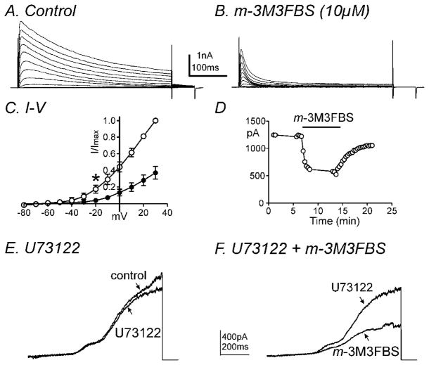
m-3M3FBS decreased IDR in a PLC-independent manner. Membrane potential was stepped from −80 to +30mV in 10mV increments from a holding potential of −80mV in (A) control conditions and (B) in the presence of m-3M3FBS. (C) Summary of normalized I-V relationships in control (○) and m-3M3FBS (●). Peak currents (I) were normalized with the peak current at +30mV (Imax). m-3M3FBS (10μM) significantly decreased IDR (n=4, * denotes P<0.05 at −20mV). All tested potentials positive to −20mV were significant. (D) Time course of inhibition of IDR generated by repetitive step depolarizations to 0mV from a holding potential of −80mV every 20 seconds before and after m-3M3FBS application. (E & F) Representative current traces are shown of ramp depolarizations stepping from −80mV to +80mV every 30 seconds in the presence of (E) U73122 (2μM) and (F) U73122 (2μM) and m-3M3FBS (10μM). m-3M3FBS decreased IDR in the presence of U73122.

