Fig. 8.
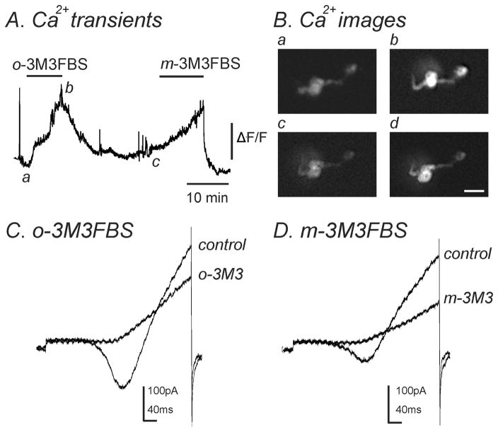
Both m-3M3FBS and o-3M3FBS increased [Ca2+]i and simultaneously decreased IL. (A) Representative trace showing an increase in [Ca2+]i upon application of o-3M3FBS (10μM) which was reversible upon washout. Addition of m-3M3FBS (10μM) to the same cell also increased [Ca2+]i which was reversible. (B) Images of a single smooth muscle cell loaded with Fluo-4AM. These images correspond to [Ca2+]i measurements shown in panel A during (a) control conditions, (b) in the presence of o-3M3FBS (10μM), (c) after washout of o-3M3FBS and (d) in the presence of m-3M3FBS (10μM). Scale bar= 20μm. (C & D) During [Ca2+]i measurements, a single ramp depolarization was applied from −80mV to +80mV over 500ms in (C) control conditions and in the presence of o-3M3FBS (o-3M3; 10μM) and (D) after washout of o-3M3FBS and in the presence of m-3M3FBS (m-3M3; 10μM). Both o-3M3FBS (n=4) and m-3M3FBS (n=7) inhibited IL.

