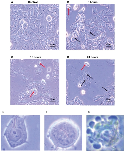
Figure 4.
Morphological characteristics of Panc-1 cells which were treated with 200 μl of Ni NWs suspension for 0 (control), 8, 16, and 24 hours and visualized with phase contrast microscope (magnification 40×). Increasing detachment of cells from the well surface is seen with increasing exposure time. (A) Adherent cells without Ni NWs (control); (B) adherent (black arrows) and suspended (red arrows) cells with internalized Ni NWs after 8 hours; (C) adherent and a greater number of suspended cells with internalized Ni NWs after 16 hours; (D) Adherent and suspended cells with internalized Ni NWs after 24 hours. (E–G) represent more specific cellular morphological changes. (E) Uptake of Ni NWs by endocytosis to the cell cytoplasm; (F) internalization of Ni NWs to the cell nucleus followed by cell shrinkage, round shaped, and initiation of membrane blebbing; (G) apoptotic body formation through final membrane blebbing.

