Figure 5.
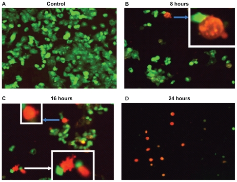
Panc-1 cells treated with Ni NWs which potentiates apoptosis. Cells were treated with 200 μL Ni NWs for 8, 16, and 24 hours and stained with ethidium bromide (EB) and acridine orange (AO) and were visualized and photographed immediately with fluorescence microscope. (A) Control; (B) 8 hours after treatment – blue arrow indicates initial membrane blebbing of the apoptosed cells; (C) 16 hours after treatment – the white arrow indicates late membrane blebbing; (D) 24 hours after treatment. Cells were stained with AO and EB where viable cells were green nuclear fluorescence and apoptotic cells exhibit a red nucleus.

