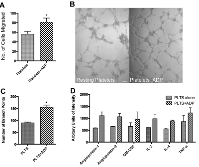
Figure 3.
Promotion of angiogenesis by ADP-stimulated platelet releasates. (A) Endothelial cell migration after exposure to the releasate from control, resting platelets (plts) or platelets activated with 25μM ADP. (B) Capillary tube formation with exposure to the releasate from resting platelets or platelets activated with 25μM ADP. The scale bar is 100μm in size. (C) Quantification of branch points generated by the releasate of platelets alone or platelets exposed to ADP. (D) Representative graph of angiogenic factors as measured by protein array that were found to have a 1.5-fold increase. * indicates P < .05.

