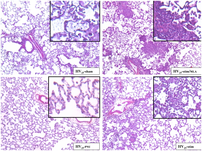
Figure 7. Lung histopathology of animals exposed to high stretch ventilation.
Hematoxylin-eosin-saffron stained sections. Original magnification of pictures x50; original magnification of insets x100. The intra-alveolar inflammation with alveolar macrophages can be seen in the HV25-sham slide and, at a lesser level, in the HV25-stim slide. Note the alveolar polymorphonuclear leukocytes in addition to the macrophages in HV25-stim/MLA group. By contrast, no intra-alveolar inflammatory cell accumulation was seen in the HV25-PNU group. In every group, the inter-alveolar walls were moderately edematous and inflamed with inflammatory mononuclear cells. The alveolar edema is here especially marked on the HV25-sham slide.

