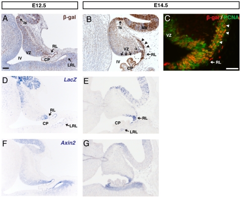
Figure 1. BAT-gal expression in the E12.5 and E14.5 cerebellum.
(A) DAB Immunohistochemistry for β-galactosidase (β-gal) on sagittal sections of E12.5 cerebellum revealed two key expression domains: the isthmus (Is) and the cerebellar rhombic lip (RL). At E14.5 (B) expression was also found in the early external granule layer (EGL, black arrowheads) but was notably absent from the ventricular zone (VZ) lining the fourth ventricle (IV). (C) Double immunofluorescence for β-gal and PCNA confirms the expression of β-gal in the RL and EGL (white arrowheads). β-gal protein was validated as a Wnt/β-catenin reporter by in situ hybridisation for LacZ (D, E) and Wnt target Axin2 (F, G)) mRNA. At both time points β-gal protein and LacZ mRNA were expressed in the same domains as Axin2, although the LacZ mRNA expression appeared less diffuse than that of Axin2. (A–B counterstained with hematoxylin. Scale bars: A, B, D–E = 100 µm, B = 50 µM).

