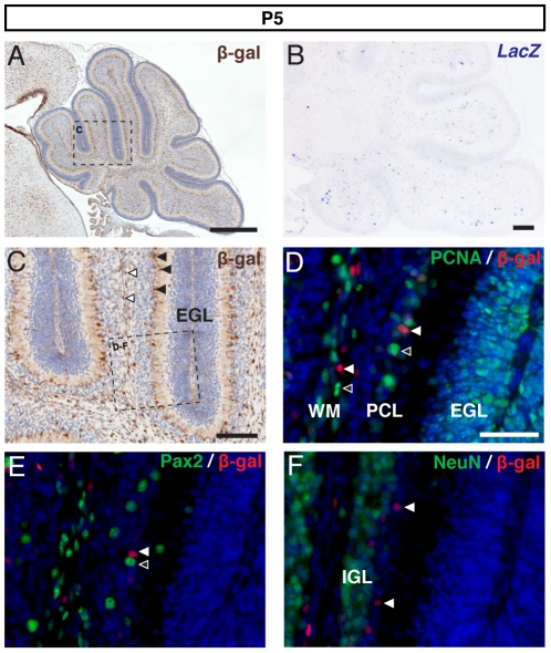
Figure 3. BAT-gal expression in the P5 cerebellum.
(A) DAB immunohistochemistry for β-gal and (B) LacZ in situ hybridisation in the P5 cerebellum. (C) Higher magnification of the region boxed in (A) reveals expression spread through all layers except the EGL. The Purkinje cell layer (PCL) and the white matter (WM) in particular contained many β-gal+ cells (black and white arrowheads respectively). Double immunofluorescence for β-gal and PCNA (D) revealed the presence of β-gal+ cells within the PCL and white matter (white arrowheads). Although β-gal+ cells were observed in close proximity to proliferating cells (unfilled arrowheads) very few β-gal+/PCNA+ cells were observed. Double immunofluorescence for β-gal and Pax2 (E) showed the close proximity of β-gal+ cells (white arrowhead) to Pax2+ interneurons (unfilled arrowhead) but no double-labelled cells were observed. Double immunofluorescence for β-gal and NeuN showed β-gal+ cells (white arrows) located outwith the IGL. (A,C counterstained with hematoxylin and D–F with Topro3. Scale bars: A = 500 µm, B–C = 100 µm, D–E = 50 µm).

