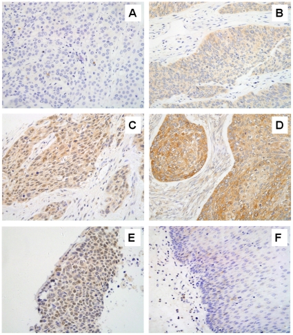
Figure 7. RAD51 is up-regulated in human esophageal cancer specimens.
Images of showing different expression level and cellular distribution are shown. (A), absence of RAD51-specific staining (blue nuclei due to Mayer's Hemalum counterstaining). (B), low levels of RAD51 in the cytoplasm, but not in nuclei, of the invasive carcinoma cells. (C), intermediate levels of RAD51 in both the cytoplasm and nuclei of the invasive carcinoma cells. (D), high levels of RAD51 in the cytoplasm, but not in nuclei, of the invasive carcinoma cells. (E), high levels of nuclear RAD51 in the nuclei of invasive carcinoma cells. No cytoplasm staining is evident. (F), absence of RAD51 staining in normal esophageal tissue. Immunohistochemistry was done as in Materials and Methods. All original images were captured at×400 magnification.

