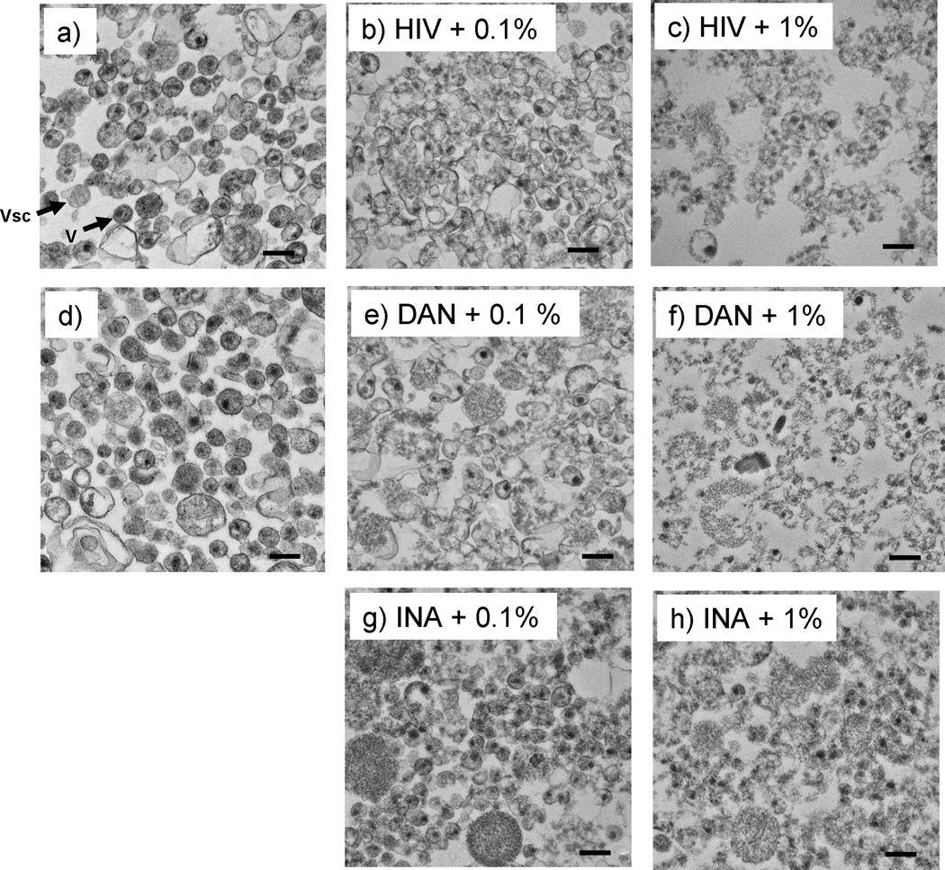
Figure 8.
Transmission electron microscopy (TEM) images of pelleted HIV after triton treatment show virions and viral protein aggregates remain after Triton treatment. Samples were treated as specified with either DAN or INA then treated with 1% Triton X-100 at room temperature for 1 hour before pelleting through 25% sucrose, and TEM analysis. a) and d) are controls of HIV and HIV+DAN+ UVA for 15 minutes, respectively; b and c are HIV controls + triton treatment with either 0.1% or 1% triton as specified; e-h are images of virus treated with DAN +UVA for 15 minutes, or INA +UVA for 15 minutes, with either 0.1% or 1% triton treatment, as specified. Arrows indicate: Vsc = microvesicle, V= HIV virion. Scale bars are 200 nm.

