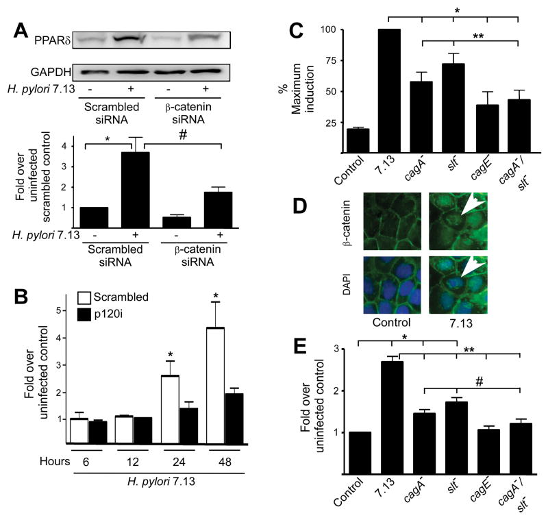
Figure 2. PPARδ expression induced by H. pylori requires β-catenin and p120 and cag island substrates. (A).
MKN28 cells transiently transfected with control or β-catenin-specific siRNA were co-cultured with strain 7.13 for 24 hours. Total protein was analyzed by Western blot using an anti-PPARδ antibody. Bar graph represents densitometric analysis of multiple experiments. Error bars, SEM for all panels. *p < .05 versus uninfected scrambled controls. #p < .05 versus 7.13-infected scrambled controls. (B) Scramble control or p120i cells were co-cultured with strain 7.13 or medium alone. Total RNA was analyzed in duplicate by real-time PCR. *p < .05 versus uninfected control. (C) MKN28 cells were transiently transfected with PPRE3-tk-luciferase and pRL-SV40, followed by infection with strain 7.13, or the cagA−, slt−, cagE− or cagA−/slt− mutants. Dual luciferase assays were then performed. *p < .001 versus 7.13-infected samples. **p < .05 versus cagA−/slt−-infected samples. (D) MKN28 cells were co-cultured with strain 7.13 or medium alone for 24 hours. Cells were stained with anti-β-catenin and AlexaFluor-488 antibodies and DAPI nuclear dye and analyzed by immunofluorescence microscopy. Arrowheads, nuclear β-catenin. (E) MKN28 cells were transiently transfected with β-catenin reporter constructs in the absence or presence of wild-type strain 7.13 or mutants lacking cagA, slt, cagE, or cagA/slt. Luciferase activity was determined 24 hours after infection. *p < .05 versus uninfected control. **p < .001 versus 7.13-infected samples. #p < .05 versus cagA−/slt−-infected samples.

