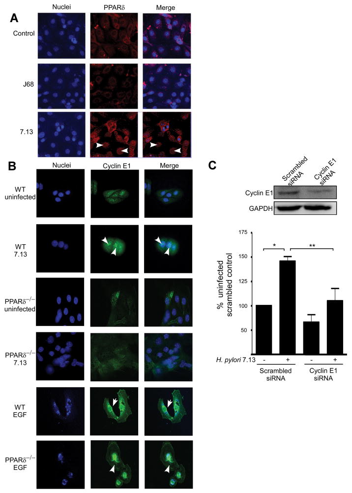
Figure 4. H. pylori induces aberrant PPARδ and cyclin E1 localization to the nucleus in gastric colonies.
(A) Primary murine gastric colonies were co-cultured with medium alone or strains 7.13 or J68 for 24 hours. Cells were incubated with an anti-PPARδ antibody, followed by an anti-goat AlexaFluor-546 antibody and TO-PRO3 nucleic acid dye and visualized by immunofluorescence microscopy. PPARδ, red; nuclei, blue. 400x magnification. Arrowheads, nuclear PPARδ. (B) Gastric colonies isolated from wild-type or PPARδ−/− mice were co-cultured with strain 7.13, EGF (10 ng/ml) or medium alone for 24 hours. Cells were incubated with an anti-Cyclin E1 antibody, followed by an anti-goat AlexaFluor-488 antibody and DAPI nucleic acid dye. Cyclin E1, green; nuclei, blue. 400x magnification. Arrowheads, nuclear Cyclin E1. (C) MKN28 cells transfected with scrambled or Cyclin E1-specific siRNA were seeded in Matrigel and infected with strain 7.13 or medium alone for 72 hours. BrdU was added to culture medium and ELISA was performed. Error bars = SEM for experiments performed on at least 3 occasions. *p < .05 versus uninfected scrambled control. #p < .04 versus 7.13-infected scrambled control.

