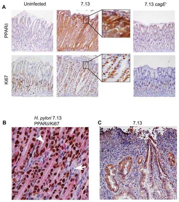
Figure 6. PPARδ and Ki67-positive epithelial cells are increased within antral gastric mucosa of Mongolian gerbils infected with wild-type H. pylori strain 7.13.
(A) Representative staining in serial sections for PPARδ and Ki67 is shown for uninfected, 7.13-infected, or 7.13 cagE−-infected gerbils. 200x magnification. (B) Dual staining for PPARδ (pink) and Ki67 (brown) is shown for H. pylori 7.13-infected gerbil. 400x magnification. Arrowhead, epithelial cell positive for PPARδ and Ki67; Arrow, stromal cell negative for PPARδ and Ki67. (C) Representative staining for PPARδ is shown in an invasive adenocarcinoma in an H. pylori 7.13-infected gerbil. 200x magnification.

