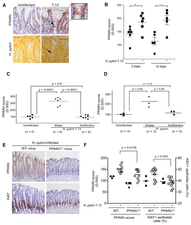
Figure 7. PPARδ expression within gastric mucosa of Mongolian gerbils, wild-type mice or PPARδ+/− mice in the presence or absence of H. pylori.
(A) Representative staining for PPARδ and H. pylori is shown for uninfected or H. pylori 7.13-infected gerbils, 3 days post-infection. 400x magnification. Arrows, cells with increased PPARδ staining adjacent to H. pylori organisms (Steiner silver stain). (B) PPARδ expression was evaluated in infected and uninfected gerbils. *, p < .05 versus uninfected samples. (C, D) Gerbils were infected with broth alone or with strain 7.13. Four weeks after H. pylori challenge, gerbils were treated with antibiotics or water and then sacrificed eight (C) or one (D) week later. (E) Representative staining for PPARδ and Ki67 is shown for H. pylori 7.13-infected wild-type and PPARδ+/− mice. 200x magnification. (F) PPARδ or Ki67 staining in gastric epithelial cells was evaluated by an observer unaware of H. pylori status or mouse genotype.

