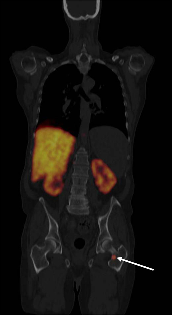
Fig. 4.
FDGal PET/CT image showing a metastasis in the neck of the left femur bone (ID27, coronal view). The liver is large and cirrhotic with no visible tumours in this plane. Radioactivity in the kidneys is physiological due to excretion of FDGal in the urine. The patient had cirrhosis (hepatitis C) and HCC was verified by biopsy. AFP was 13 ng/ml.

