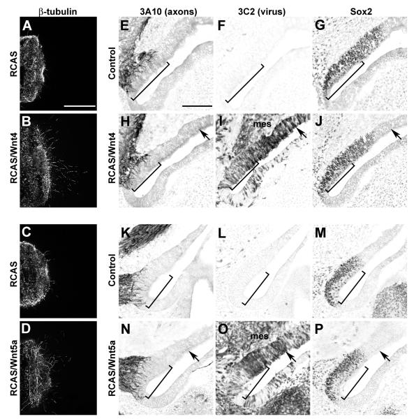
Fig. 6. Spinal cord axons but not inner ear SAG axons are responsive to Wnt4 and Wnt5a delivered via RCAS retroviral vectors.
Confocal images of E6 spinal cord explants cultured for 24h in the presence of supernatant collected from virus-infected cells (A-C). Scale bar = 200 μm. More axons emerge from spinal cord explants grown in culture supernatant collected from fibroblasts infected with RCAS/Wnt4 (A) or RCAS/Wnt5a (B) as compared to RCAS (C) or supernatant from uninfected fibroblasts (not shown). Infection with Wnt4 or Wnt5a viruses on E3 does not affect innervation to the saccular macula on E6 (E-P). Serial horizontal sections through inner ears were immunostained with antibodies to detect axons (3A10), virus (3C2) or prosensory domains (Sox2; E-P). Images are oriented with medial to the left and anterior down. Images of the right ears were flipped to mimic the orientation of the left ears to faciliate comparisons. Scale bar=100 μm. Virus-mediated misexpression of Wnt4 (H-J) or Wnt5a (N-P) does not alter the trajectory of SAG axons into the prosensory domains of the E6 inner ear when compared with the contralateral uninfected control sides (E-G and K-M, respectively). The innervation density (brackets) of Sox2-positive prosensory maculae is similar on control and experimental sides, while nearby virus-infected non-sensory patches (black arrows) remain free of axons. Furthermore, axons are not attracted into infected mesenchyme (mes).

