Figure 7.
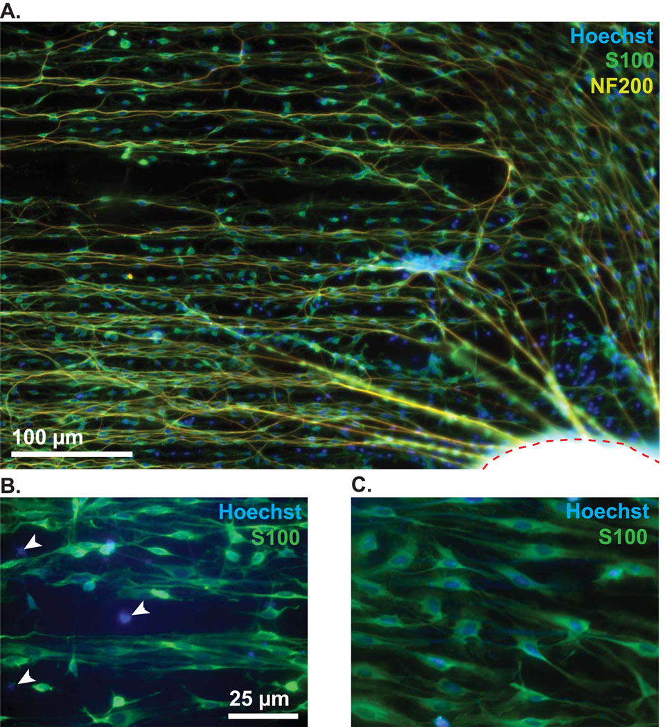
Spiral ganglion explant on patterned HMA/HDDMA polymer. (A) The explant body is outlined with a dashed red line on the bottom right corner. NF200-labeled neurites (yellow) extend initially from the explant in a radial fashion and are subsequently induced to turn parallel to the pattern which is oriented along the horizontal plane. Anti-S100 antibody (green) and Hoechst (blue) were used to label SCs and nuclei respectively. Notably, there are several S100-negative cell nuclei clustered close to the explant, as exemplified in the higher magnification images below. (B) White arrowheads indicate S100-negative cell nuclei. (C) Image taken further away from the explant (~ 200 µm) demonstrating that all nuclei are associated with S100-positive cells. The micropatterned MAs support SC outgrowth more favorably than S100-negative cells such as fibroblasts.

