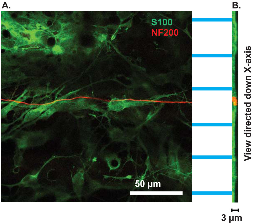
Figure 9.
SGN neurite grow within the microgrooves of patterned HMA/HDDMA. (A) Section of confocal z-stack from SG culture immunostained with anti-S100 (green) and anti-NF200 (red) antibodies. The neurite grows within the groove, demonstrated as a cellular stripe between acellular ridges. Scale bar=50 µm. (B) Ninety degree rotation of the stack to allow viewing down the x-axis. The grooves are evident as the regions with thicker S100 labeling compared with the ridges. The neurite (red) remains confined to the groove region. Scale bar=3 µm.

