Fig. 1.
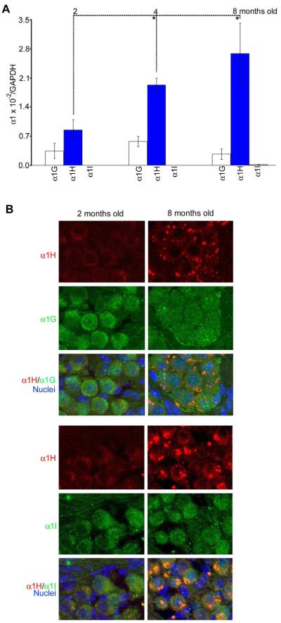
Age-related changes in expression of the α1 subunits in the cochlea during aging. (A) Mean (+SD) expression of the α1G, α1H and α1I subunits as determined by real-time RT-PCR quantification in female mouse cochleae at 2, 4, and 8 months of age (n=6 per group), and a significant difference was found between adjacent ages for the α1H gene (t-test, *p < 0.01). The expression level for each subunit was determined by the ratio between each α1 subunit and GAPDH to eliminate possible differences in the amount of total RNAs from each sample. (B) Immunocytochemistry showing co-localization of the α1H subunit with the other two subunits in SGNs at 2 and 8 months of age.

