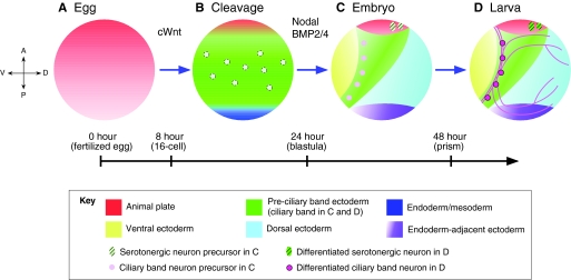
Fig. 4.
A four-step model for specification and organization of the sea urchin embryo nervous system. (A) In the first step, an anterior neuroectoderm regulatory state (red) is present throughout the egg and much of the embryo during early cleavage stages. (B) In the second step, which occurs during very early blastula stages, this state is eliminated by canonical Wnt (cWnt)-dependent signals from all but the anterior neuroectoderm, revealing a ciliary band-like neuroectoderm (green) that contains scattered neural precursors (light pink circles). (C) In the third step, which occurs during the mesenchyme blastula/early gastrula stages, Nodal and BMP2/4 signals convert ventral and dorsal ectoderm to non-neural ectoderm except in the anterior neuroectoderm (red) and ciliary band (green), which are protected from these signals. (D) During the fourth and final step, by which point the embryo has transitioned into a larva, CBE and ANE neural progenitors differentiate. The timeline indicates hours post-fertilization and embryonic stages.

