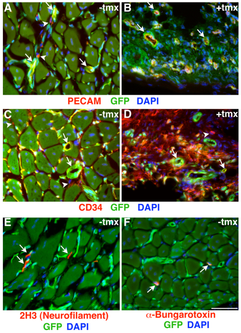
Fig. 4.
Robust vascularization of EDL muscles transplanted with or without prior ablation of Pax7+ cells and innervation of control EDL muscle grafts. (A-F) Fluorescent microscopy of Pax7+/CE;R26ReGFP-DTA/lacz EDL grafts without (A,C,E,F) and with (B,D) exposure of the donor EDL muscle to tamoxifen (tmx) prior to transplantation. (A,B) PECAM is pseudocolored in red, GFP in green and DAPI in blue. White arrows indicate PECAM+ capillaries; white arrowheads indicate GFP–/PECAM+ host infiltrating cells. (C,D) CD34 is pseudocolored in red, GFP in green and DAPI in blue. White arrows indicate CD34+ capillaries; white arrowheads indicate GFP–/CD34+ host infiltrating cells. (E) 2H3 (neurofilament)-stained neuronal axons are pseudocolored in red, GFP in green and DAPI in blue. White arrows indicate 2H3+/GFP– neuronal processes. (F) Alexa568-conjugated α-bungarotoxin-labeled motor end plates (via binding to nicotinic acetylcholine receptors) are pseudocolored in red, GFP in green and DAPI in blue. White arrows indicate bungarotoxin+/GFP– motor end plates. Scale bar: 50 μm.

