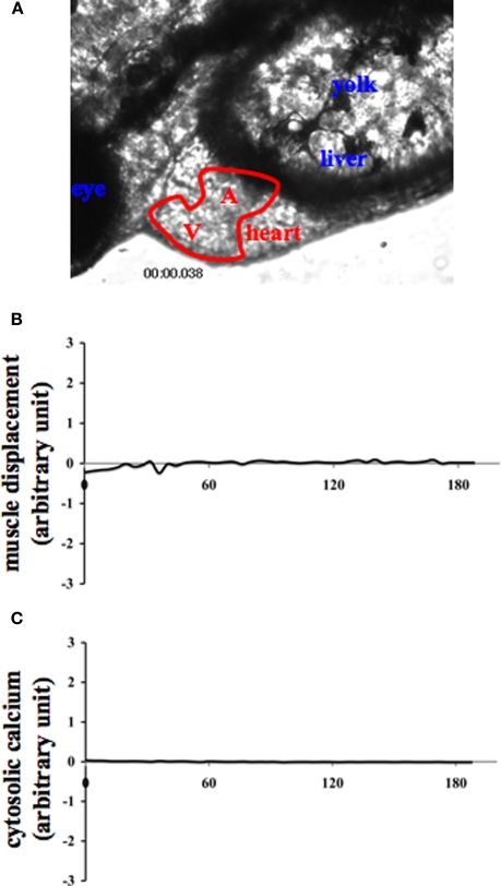
Figure 2.
Internal muscle movement and cytosolic calcium are studied in various organs of zebrafish. Internal organs of zebrafish were indicated by the corresponding text (A). A and V denote the atrium and ventricle of the heart, respectively. To verify the specificity of muscle contraction and calcium oscillation in the heart and to avoid any movement artifact, changes in muscle movement (B) and cytosolic calcium (C) of the corresponding fish were also measured in the eye. The eye measurement can therefore represent an internal negative control for cytosolic calcium recording.

