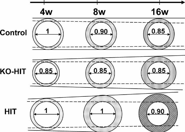
Fig. 6.

Schematic illustration of bone-growth patterns in mice with altered tissue and/or serum IGF-1 levels. Female control mice show a slight increase in total cross-sectional area during the first 8 weeks of life. Later on, a marked increase in cortical bone area and TMD, associated with marrow in-filling, allows an efficient structure to support the age-related increase in body weight. KO-HIT female mice exhibit a significantly smaller total cross-sectional area at 4 weeks, but at 8 weeks they show a “catch up” growth and do not differ significantly from controls. Nonetheless, between 8 and 16 weeks, KO-HIT female show a significant increase in total cross-sectional area associated with a marked increase in cortical thickness and no changes in marrow area, suggesting increased periosteal bone apposition. In contrast, HIT mice show significant increases in both total cross-sectional area and cortical bone area between 4 and 8 and 8 and 16 weeks of age. These increases were associated with a decrease in marrow area, suggesting that both periosteal and endosteal bone apposition took place.
