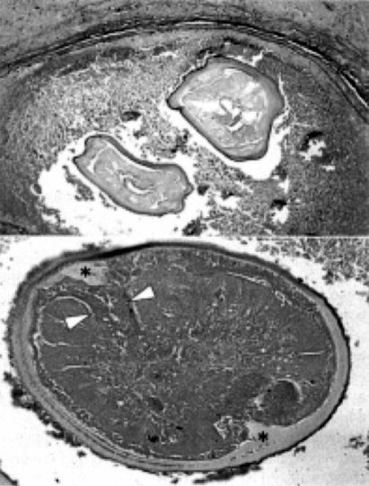
Fig. 3.
Histopathologic findings of the nodule. Two transverse sections of an immature worm of D. immtis are seen in a small pulmonary artery (upper, Elastica van Gieson stain), and a transverse section of an immature adult worm showing large lateral chords (arrow head) with internal longitudinal ridges (*) and multilayered cuticle (bottom, HE stain).

