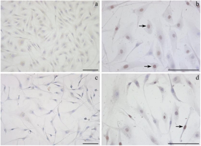
Figure 4.
Representative photographs of selected fields of TUNEL-stained ear fibroblasts are shown. The TUNEL-positive nuclei, indicating apoptotic cells, are stained brown (black arrows), while the vital nuclei are stained violet (haematoxylin). (a) Ear fibroblasts in culture for 24 h (no OTA). (b) Ear fibroblasts treated with 0.6 μg/mL OTA for 24 h. (c) Ear fibroblasts treated with 1.25 μg/mL OTA for 24 h. (d) Ear fibroblasts treated with 1 nM α-tocopherol and 0.6 μg/mL OTA for 24 h. Bars = 200 μm.

