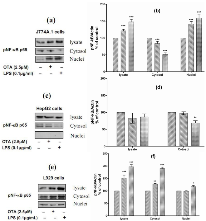
Figure 5.
Panels (a), (c), and (e) Representative Western blots with antibodies detecting phospho-NF-κB p65 in the cell lysate, cytosolic and nuclear fractions after incubation with vehicle, 2.5 µmol/L OTA, or 0.1 µg/mL LPS up to 24 h. Panels (b), (d), and (e) Densitometric analysis of samples obtained from cells treated with vehicle ( ), 2.5 µmol/L OTA (
 ) or 0.1 µg/mL LPS (
 ). Panel (a) and (b) obtained from J774A.1 cells, Panel (c) and (d) from HepG2 cells, Panels (e) and (f) from L929 cells. Data obtained from three independent experiments, which presented as percent of control (mean ± SEM) after calibration for actin density. Marked columns were statistically different from controls (* P < 0.05; ** P < 0.01; *** P < 0.001)(The Role of NF-κB in OTA/ LPS mediated TNF-α release).

