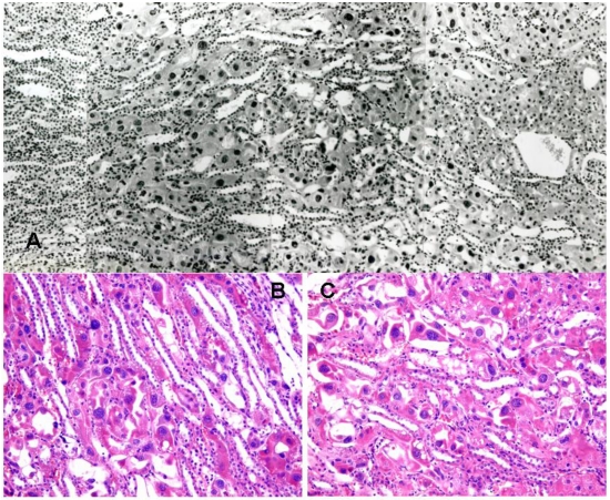
Figure 1.
Rat renal histopathology (stained with H & E) after five weeks on a P. polonicum-contaminated diet (rat 4). A, part of saggital section traversing from innermost glomerulus in cortex (right) into the medulla, showing extensive, diffuse karyocytomegaly across the region. B and C, two representative examples in the region associated with concentration of P3 segments of nephrons, showing detail of enlarged nuclei in distorted cells.

