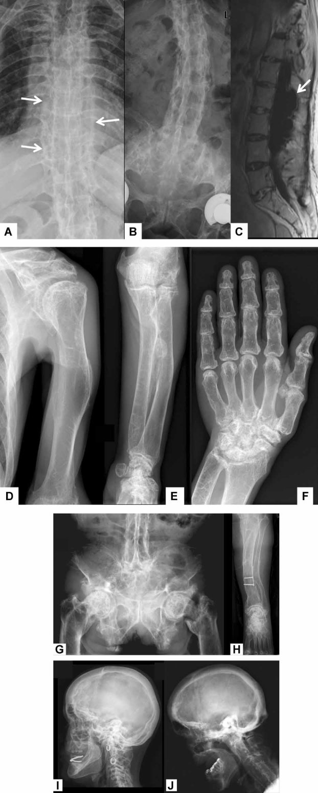
Fig. 3.

Radiographic findings in the two ARHP patients with the homozygous DMP1 mutation. Calcifications (arrows) of the spinal ligaments in the thoracic (A) and lumbar spine (B) in patient 1. Spinal MRI of patient 1 showed a large dural ectasia (arrow) in the midthoracic and lumbar spine (C). Abnormal bone structure and short and deformed long bones (D, humerus; E, radius and ulna; F, hand; H, tibia and fibula) in patient 1. Patient 2 had a pathologic fracture of the left femoral neck (G). Both patients had significant cranial hyperostosis (I, patient 1; J, patient 2).
