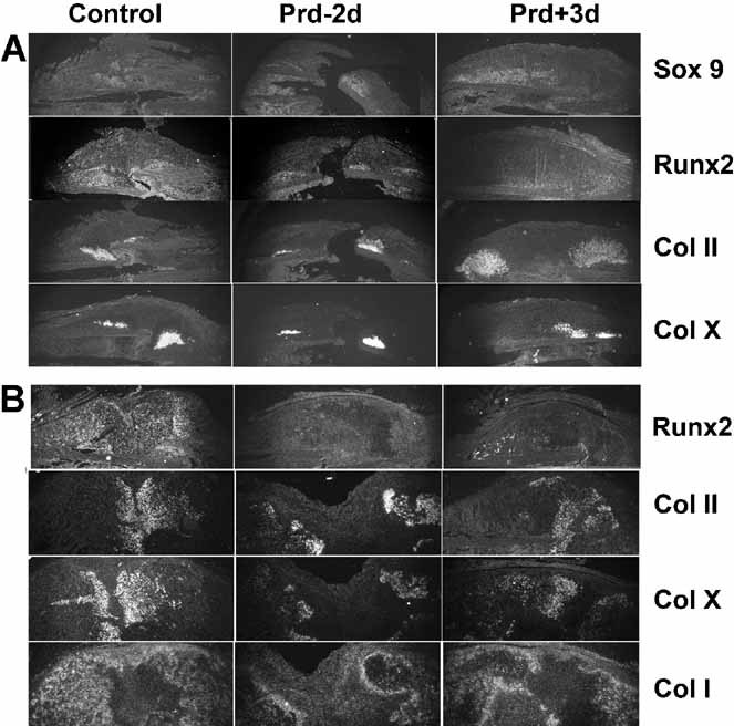
Fig. 4.

In situ hybridization analyses. Sections from the calluses of control mice and mice on the PRD were subjected to in situ hybridization analyses for the mRNAs indicated on the right. Data in A were obtained 7 days after fracture, whereas those in B was obtained 14 days after fracture. The upper part of each image represents half of the callus, with the original cortical bone on the lower aspect. Data represent those obtained from two sections from each of three mice for each time point and dietary condition.
