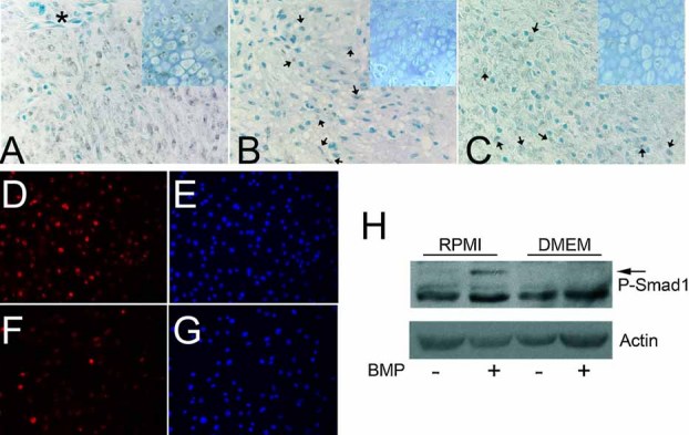
Fig. 6.

Phosphate restriction attenuates nuclear localization of pSmad1. (A) Control callus. Most stromal cells are positive for nuclear pSmad1 (brown stain); asterisk indicates a blood vessel with no nuclear pSmad in endothelial cells. (B, C) Calluses of mice placed on the PRD 2 days before or 3 days after fracture. Arrows point to the rare cells with nuclear pSmad1. Insets in the right upper corner represent chondrocytes from the same calluses. (D–H) ST2 cells were cultured in RPMI (5.6 mM phosphate; D, E) or DMEM (1 mM phosphate; F, G). Immunocytochemistry for pSmad was performed 90 minutes after rhBMP-2 (D, F). DAPI staining was performed to permit identification of nuclei (E, G). (H) Phosphate restriction attenuates Smad phosphorylation. Cells were cultured as earlier, and lysates were subjected to Western blot analyses for pSmad (arrow). Data are representative of those obtained in three independent experiments.
