FIGURE 5.

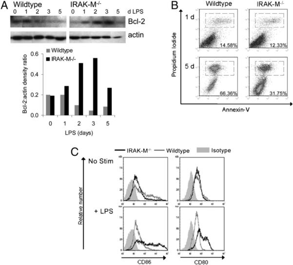
IRAK-M−/− DCs live longer than WT DCs. IRAK-M−/− and WT DCs were cultured in CellGenix media without growth factors plus 50 ng/ml LPS. A, At the times indicated, cells were harvested, lysed with buffer containing protease and phosphatase inhibitors, and subjected to Western blot. Blots were probed for Bcl-2, stripped, and reprobed for actin; densitometry is also shown (lower panel). Abs used are listed in Materials and Methods. B, At the times indicated, cells were harvested and stained for Annexin V and PI; numbers indicate the percentage of gated (dead) cells. Data are representative of four independent experiments. C, After 5 d of the indicated stimulus, cells were harvested, stained, and analyzed by quantitative flow cytometry. Plots are gated on viable cells and shown as nonnormalized to indicate the relative number of events.
