
Fig. 2.
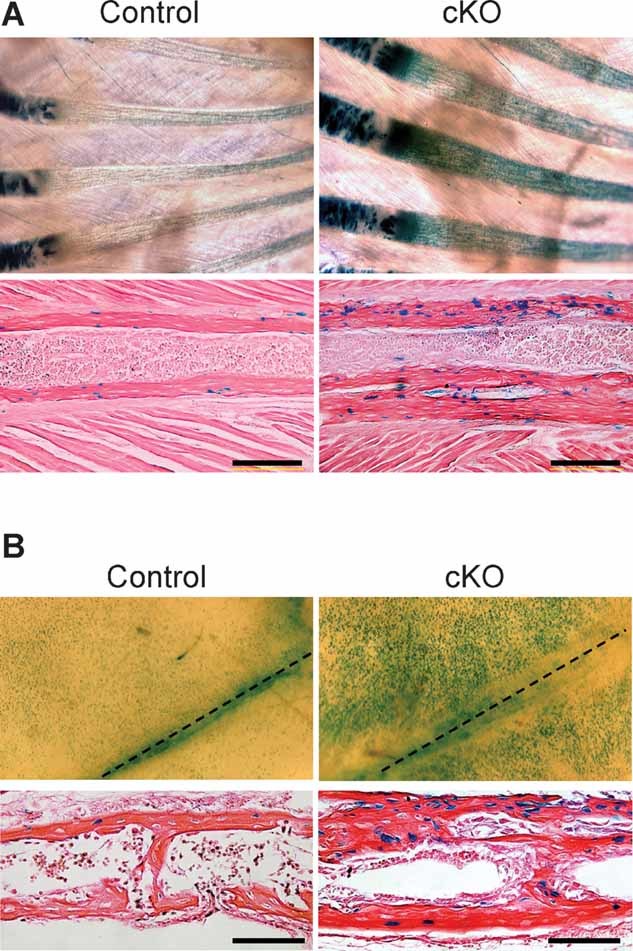
Upregulation of canonical Wnt signaling in Bmpr1a cKO mice. (A) Canonical Wnt signaling in P14 rib bones assessed using TOPGAL mice. Whole-mount β-gal staining showed increased staining in cKO rib bones compared with controls (upper panels). Histologic analysis showed an increased number of β-gal-positive osteoblasts in cKO rib bones compared with controls (lower panels). Bar: 200 µm. (B) Canonical Wnt signaling in P10 skull bones assessed using TOPGAL mice. Whole-mount β-gal staining showed increased staining in cKO calvariae compared with controls (upper panels). Histologic analysis showed an increased number of β-gal-positive osteoblasts in cKO calvariae compared with controls (lower panels). Dotted lines indicate the sagittal suture. Bar: 100 µm.