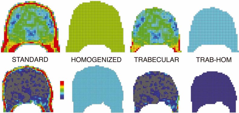
Fig. 1.

To illustrate the biomechanical effects of spatial variation in bone density, transverse cross sections of the finite-element model of a vertebra are shown for a subject with relatively strong bone (top row) and one with relatively weak bone (bottom row). Each finite element is assigned material properties based on vBMD data from the QCT scan for that element, ranging from high-density (red) to low-density (gray) bone. For each subject, cross sections are shown for four models: the unaltered vertebra (“STANDARD”); the vertebra with all elements assigned the average vBMD value for that model (“HOMOGENIZED”); a model consisting only of the trabecular compartment, in which the outer 2 mm of bone is virtually removed (“TRABECULAR”); and a homogenized version of that trabecular model with all elements assigned the average vBMD of the trabecular compartment (“TRAB-HOM”). In each case, the resulting finite-element models are virtually compressed to failure to estimate compressive strength, resulting in multiple strength outcomes for each subject.
