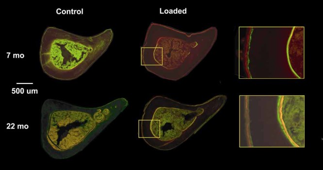
Fig. 2.

Fluorescent photomicrographs of mid-diaphyseal tibial sections from loaded and control tibias of 22- and 7-month-old mice. Samples were collected on day 11 following tibial compression on days 1 through 5 and fluorochrome labeling on days 5 (green) and 10 (red). An increase in endocortical and periosteal labeled surface is evident in loaded tibias of both age groups compared with controls. (Shown are tibias from the 10-N load group.)
