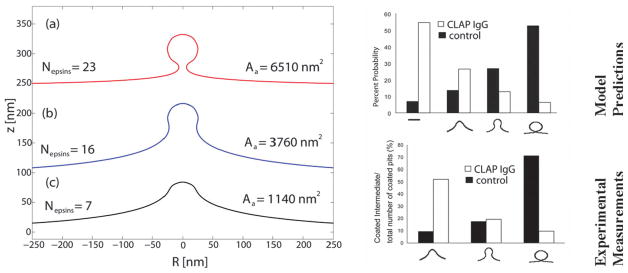
Fig. 4.
(Reproduced with permission from ref. 125) Left: membrane deformation profiles under the influence of imposed curvature of the epsin shell model for three different coat areas, here κ = 20 kBT. For the largest coat area, the membrane shape is reminiscent of a clathrin-coated vesicle. Right: calculated (top) and experimentally measured (bottom) probability of observing a clathrin-coated vesicular bud of given size in WT cells (filled) and CLAP IgG injected cells (unfilled).

