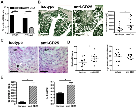
Figure 5. In vivo ablation of CD25+ cells impairs regulation of colonic granulomas and antigen-specific Th2 responses.
A) Percentage of CD25+ or CTLA-4+ mLN lymphocytes from mice with chronic infection after treatment with anti-CD25 mAb, or isotype control. Antibodies given at 2 week intervals from wk 9 to wk 13, tissues sampled at wk 14. Data are mean % positive (± SEM). B) Photomicrographs of colonic tissue and quantification of granuloma areas after antibody treatment. Scale bar = 200 µm; egg denoted ‘Sm’. Data is granuloma area (µm2) at wk 14 calculated from 3-4 separate histological sections per individual animal (n = 4). C) Colonic granulomas (x100) stained with H&E showing eosinophils (closed arrow) and large mononuclear cells (open arrow) in isotype mAb treated (left) and anti-CD25 treated (right|) infected mice. Scale bar = 200 µm; egg denoted ‘Sm’. D) Numbers of eosinophils (left) and large mononuclear cells enumerated from the sections above; Bars are mean cell counts / group (n = 4) with 3–4 fields of view / mouse. E) Antigen-specific mLN responses in anti-CD25 mAb treated mice; data are means of cpm 3H-thymidine incorporation and IL-4 secretion pg/ml (n = 3).

