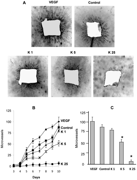
Figure 3. Kahweol inhibits endothelial cell sprouting from aortic rings in a dose-dependent manner.
Aortic ring assay was performed as described in Materials and methods. A) Negative of photographs (x20) of aortic rings (lateral view) after 10 days of incubation in a 3D collagen gel overlayed with complete medium in the presence of 20 mg/mL VEGF, 0.05% DMSO (the vehicle taken as a control), or kahweol at 1, 5 and 25 µM (K1, K5, K25, respectively). Experiments were repeated at least three times. B) Microvessel time course for the different treatments mentioned in A. Data are given as microvessel total count at different incubation times (spanning from 3 to to 10 days), and they are means±S.D. of three different experiments. C) Microvessel total count after 10 days of incubation. Data are given as microvessel total count, and they are means±S.D. of three different experiments. *Statistically significant (p<0.01) as compared to control values, according to a two-tailed Student's t-test.

