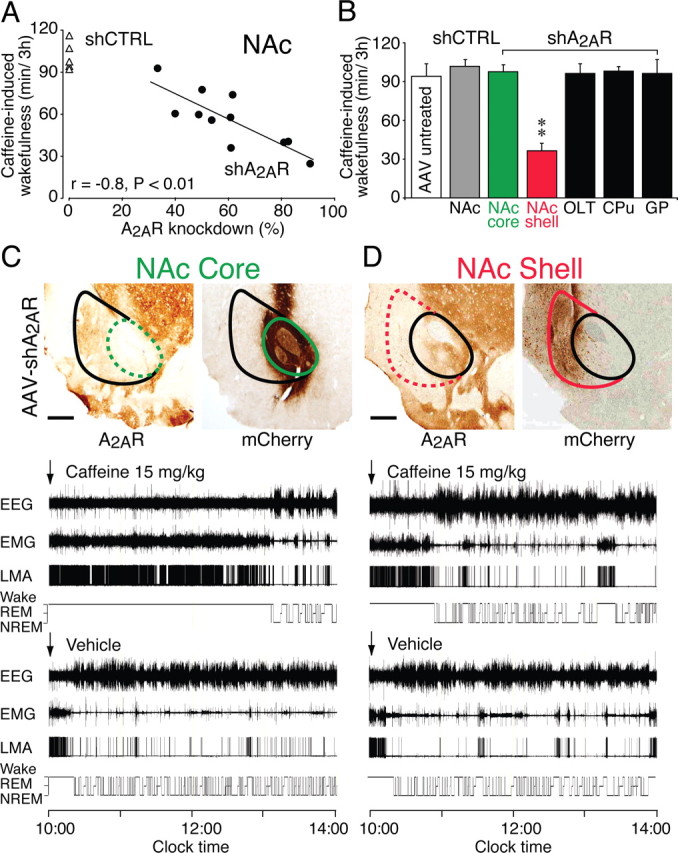
Figure 5.

Arousal effect of caffeine was abolished in rats with site-specific deletion of A2ARs in the shell of the NAc. A, The loss of the arousal response to caffeine (15 mg/kg, i.p.) in rats that had received AAV–shA2AR injections (black circles) into the NAc correlated closely with the knockdown of A2ARs in the NAc, whereas there was very little variation in caffeine-induced wakefulness in control animals injected with shCTRL (white triangles). B, Caffeine-induced wakefulness for 3 h in AAV-untreated rats and in rats that received injection of AAV–shCTRL into the NAc or injection of AAV–shA2AR into the NAc shell and core, OLT, CPu, and GP. Caffeine was given intraperitoneally at 15 mg/kg. C, D, Typical sections from two rats injected bilaterally with AAV carrying shA2AR into the shell or core of the NAc that were stained with mouse monoclonal antibody against A2AR showed dominant depletion of A2ARs either in the shell (D, right photomicrograph) or the core (C, left photomicrograph) of the NAc. Adjacent sections to C and D were stained with rabbit polyclonal antibody against mCherry (Clontech) to confirm the extent to which neural cells in the NAc were transfected with AAV–shA2AR (C, D, right photomicrographs). The red circles in the photomicrographs in D outline the entire NAc, including both the core and shell region, whereas the green circles in the photomicrographs in C delineate the core of the NAc. The polysomnographic recordings in C and D show typical examples of EEG, EMG, LMA, and hypnograms after administration of caffeine at a dose of 15 mg/kg (top polysomnographic panel) or vehicle (bottom polysomnographic panel) in two rats with AAV–shA2AR infections of the shell (D) or the core (C) of the NAc. Data are presented as mean ± SEM (n = 5–6 per AAV-treatment). **p < 0.01 compared with AAV-untreated or AAV–shCTRL-injected rats, assessed by one-way ANOVA. Scale bars: C, D, 300 μm.
