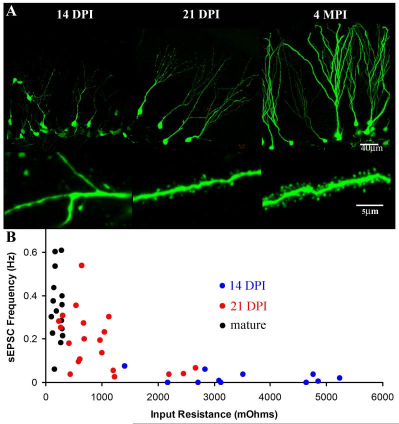
Figure 1.
The Integration of Newborn Neurons into the Adult Dentate Gyrus.
(A). The morphology of newborn neurons is depicted by labeling with a retrovirus expressing EGFP using the ubiquitin promoter (pRubi) in 6 to 8 week old mice. At 14 days post-injection (DPI) the aspiny dendrites (lower panel) have reached the inner molecular layer (upper panel). Only 1 week later at 21 DPI the majority of dendrites span the molecular layer (upper panel) and have numerous dendritic spines (lower panel). At 4 months post-injection (MPI) the neurons are fully mature with elaborate dendritic arbors (upper panel) and numerous mature dendritic spines (lower panel). (B) The stages of neuronal maturation depicted above can easily be discerned by the electrophysiological profile of the cells as well. Each point represents a single whole-cell recording of spontaneous excitatory post-synaptic currents (sEPSC) frequency plotted as a function of cellular input resistance. The sEPSC frequency correlates to the density of excitatory synaptic inputs onto the cell and the input resistance of the cell decreases as the size of the cell increases. At 14 DPI there is high input resistance and almost no excitatory synaptic currents. By 21 DPI there is a decrease in the input resistance and an increase in the sEPSC frequency. Mature neurons have the lowest input resistance and the greatest frequency of sEPSCs.

