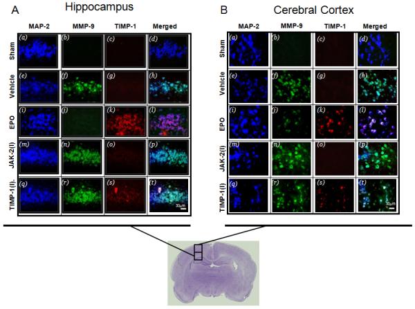
Figure 7. TIMP-1 and MMP-9 expression in the brain of neonatal hypoxic ischemic rats after inhibition of JAK-2 and TIMP-1.

The expression of TIMP-1 (Texas Red) and MMP-9 (green, FITC) in neurons (blue, AMCA) in the (A) hippocampus and (B) cortex of neonatal HI animals treated with (5U/g) EPO. Merge of TIMP-1 and MMP-9 with and without EPO are shown in the right panes. The co-localization of TIMP-1and MMP-9 in neurons 48 hours after hypoxia ischemia in EPO treated neonatal rats. Both inhibitors suppressed the upregulation of TIMP-1 in the presence of EPO treatment.
