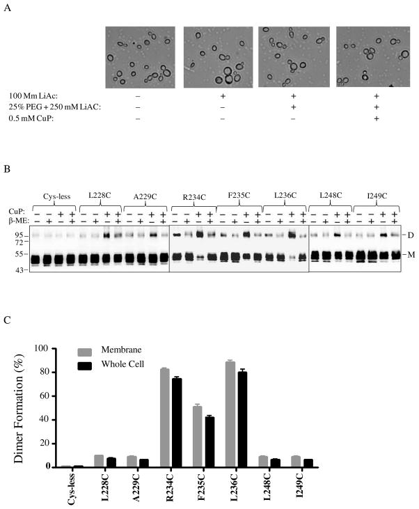
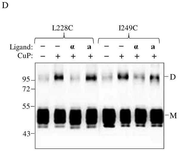
Figure 4.
Analyses of Ste2p dimerization in whole yeast cells. Cells expressing various Ste2p IL3 cysteine mutants were permeabilized and exposed to CuP. A: Images of cells treated with various reagents used during the whole cell disulfide cross-linking CuP treatment. B: Blots of seven IL3 Cys mutants selected for whole cell cross-linking evaluation. The samples were probed with anti-FLAG antibody to detect the presence of Ste2p at either the monomer (M) or dimer (D) positions. Each preparation was untreated (lanes −, −) or treated with β-ME (lanes −+) or CuP (lanes +, −) or with CuP and β-ME (lanes +, +). C: A graph comparing the percentage of the Ste2p mutant receptors that formed crosslinked dimers in membrane preparations and whole cells. D. Whole cells expressing receptors L228C and I249C were untreated or incubated with ligand prior to Cu-P treatments. The blots were probed with anti-FLAG antibody to detect the presence of Ste2p at either the monomer or dimer positions, respectively. Each preparation was untreated (lanes −, −) or treated with Cu-P (lanes −, +) or with α-factor and Cu-P (lanes α, +) or with α-factor antagonist and Cu-P (lanes a, +).


