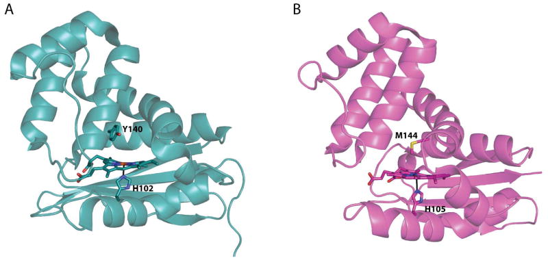
Figure 1.
Structures of the (A) O2-binding WT Tt H-NOX (1U55, chain A) in teal (15) and (B) a non-O2-binding H-NOX from Nostoc in magenta (14). Hemes and coordinating histidines as well as the distal pocket residue about the heme are shown in sticks. A global alignment of these two structures results in an RMSD of 1.88 Å (using Cα − Cα).

