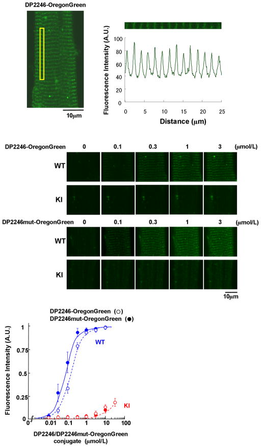
Figure 6.
Localization and the characteristics of DP2246 binding to the cellular components (SR in situ) of saponin-permeabilized WT and KI cardiomyocytes. (Top) The DP2246, fluorescently labeled with Oregon Green 514 (Molecular Probes, OR), was delivered into the cardiomyocytes. (Middle) Representative images of Oregon Green-labeled DP2246 or DP2246mut in WT and KI cardiomyocytes. (Bottom) The plot of the intensity of the fluorescence along the sarcomeres as a function of the concentration of DP2246 (open circle) /DP2246mut (closed circle)-Oregon Green. Data represent means±SE of 4 to 5 cells from 3 to 4 hearts.

