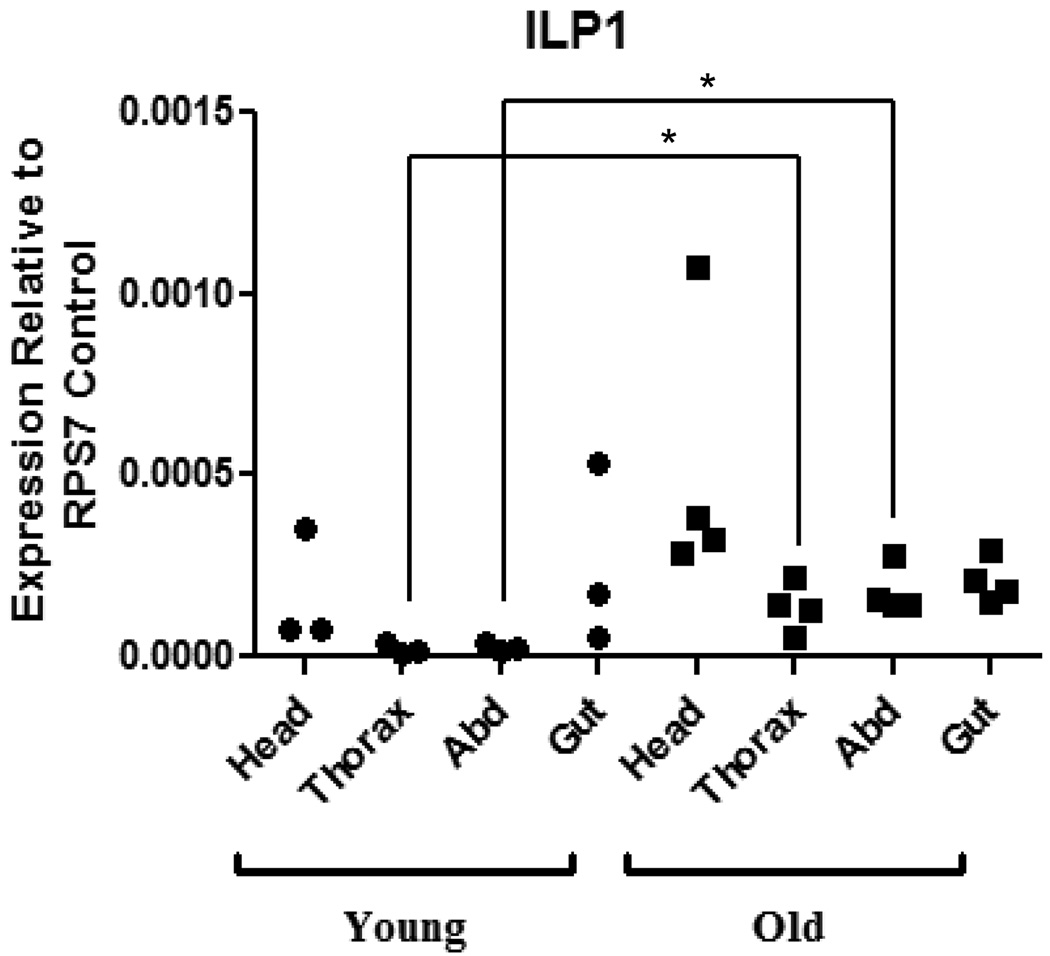
Figure 2. AnsILP1 expression is increased in the thorax and abdomen of older An. stephensi females.
Total RNAs from the head, thorax, abdominal body wall (Abd), and gut tissue (midgut, hindgut, and Malpighian tubules) of females were isolated on days 3–4 (Young) and days 15–16 (Old) post-emergence. AnsILP expression in each tissue was quantified by qRT-PCR. Expression levels are shown relative to expression of ribosomal protein S7 (RPS7) in the same sample. For statistical analysis, data were log-transformed and analyzed by ANOVA followed by Newman-Keuls post-test (α = 0.05).

